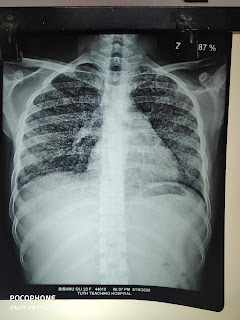
chest x-rays

Miliary TB fig 1 
Here is a chest x-ray of a young girl with Factor X deficiency from rural Rolpa. The disease takes away the zeal of life. she is a perfect example of how disease torments continuously. she was admitted multiple times with bleeding from her orifices. she even had a rupture of ovarian cyst and hemoperitoneum. when she presented last time, she had an acute onset of shortness of breath and fever for 10 days. her fever had subsided while she was still short of breath. her covid 19 pcr was negative. initially, her chest x-ray and symptoms corroborated with the current covid 19 viral infection. Her brother was short of money and tried to take her away against medical advice. however, we managed to get free bed for her and convinced him to stay back in hospital. when she had very delayed improvement than expected in her oxygen requirement and respiration, we sent covid 19 PCR again and kept her on steroid. we repeated chest x-ray and it revealed miliary shadows suggestive of miliary tuberculosis. we prescribed her antitubercular treatment and then discharged her. fortunately, we could hold her back. or else she would die of miliary TB. 


fig.2 cannonball metastases 
Here is a chest x-ray of a young beautiful lady who comes with a history,decreased appetite,and significant unintentional weight loss for a month. she was totally stable.Her vitals were all fine. on digging her past, she had an abortion at about 3 months 1 year back. she had no localizing symptoms elsewhere in body. chest x-ray had malignant look suggestive of metastases. the cannon ball appearances on chest x-ray in such a young lady could be from breast. But she had no mass or lumps there. She had no peripheral lymph nodes. It was difficult to disclose our suspicion of malignancy to her. we asked her to bring Contrast CT chest on next opd. I didn't get chance to meet her. Following her CT chest on radiology department, report was suggestive of choriocarcinoma. Bad x-ray, stable patient. Sinister is the cause. 


Fig 3: ....................  


A 67-year-old female, former smoker came to an emergency with an increase in shortness of breath, abdominal distension, and leg swelling. She was managed with diuretics and was discharged from emergency observation on the next day. Basic investigations and echocardiography were done. she was managed in line with COPD and was discharged. She came to cardiology opd in the mid of the day just at the time to have a tea break. I was on a hurry. I saw her echo report which mentioned dilated RA, RV, and moderate TR. Assuming the diagnosis of COPD, I nearly dispatched the case. However, she had the use of her sternocleidomastoid muscles. On asking her symptoms, she had fatigue and exertional SOB for months and had a history of Pulmonary TB about 15 years back. when we saw her chest x-ray, we considered repeat echocardiography. my senior took her to the echo room and did an echo on her. He found the features suggestive of constrictive pericarditis. The pericardial calcification in a chest x-ray is highly suggestive of constrictive pericarditis. besides, when the edema is less prominent in right-sided heart failure symptoms, ascites praecox is a possibility. So, don't see a patient in a hurry.  

                                                    fig.4 : right middle zone consolidation 

A recently retired old man, smoker with no other past comorbidities came to OPD with a history of right-sided pleuritic chest pain for 2 days. No history of fever. He had a dry cough at that time. However, he gave a history of right leg swelling. basic investigations are done. Complete blood count, renal function, liver function were normal.  He was prescribed antibiotics after seeing the very X-ray chest. what did we miss here? 
The right leg swelling should have been taken quite seriously. Unilateral swelling of the leg with chest pain should prompt the suspicion of thromboembolism. 
venous doppler of the legs would be a better approach in such a case. he had no other features suggestive of pneumonia. No leucocytosis, no fever. 
Remember, pulmonary embolism is a great masquerade in clinical practice. It can mimic heart failure , pneumonia in elderly populations. 


Comments

Popular posts from this blog

अन्नपूर्ण बेस क्याम्पको यात्रा आत्मसन्तुष्टि वा अहम् सन्तुष्टि !!

बाटो बिराएका रोगहरु ।

नेपाल पूर्वको यात्रा , सिकाउँछ पाठ धेरै ।